Atherosclerotic disease occurs when plaque builds up within the walls of arteries. Plaque contains cholesterol, calcium, and other materials. Over time, this buildup can harden and narrow the arteries, reducing the flow of oxygen-rich blood.
Atherosclerosis can affect arteries throughout the body, including those in the neck and those supplying the brain and heart. If plaque grows, ruptures, or causes a blood clot, it can lead to serious complications such as stroke or heart attack.
Treatment depends on the location and severity of the disease. It may include lifestyle changes, medicines to control cholesterol or blood pressure, and, in selected cases, a procedure to restore blood flow.
Carotid stenosis most often develops where the main carotid artery divides in the neck. Plaque at this point can limit blood flow or send debris into arteries that supply the eye and brain. Some patients have no symptoms. Others are diagnosed after a TIA, stroke, or brief loss of vision in one eye.
Carotid ultrasound is often the first test. It measures blood-flow speed and estimates the degree of narrowing without radiation or contrast dye. CT angiography or MR angiography gives a fuller view of the plaque and circulation beyond it. Catheter angiography may be used when the team needs more detail or is considering a catheter-based treatment.
Every plan includes control of stroke risk factors. Care may involve antiplatelet medicine, cholesterol treatment, control of blood pressure and diabetes, help quitting tobacco, exercise, and diet changes. Patients should take these medicines only as directed by their clinicians.
Opening the artery can lower future stroke risk in selected patients with severe narrowing, especially after symptoms. Carotid endarterectomy removes plaque through an incision in the neck. Carotid stenting uses a small mesh tube to widen the artery. The team chooses an approach based on the patient's health, artery shape, and procedure risk.
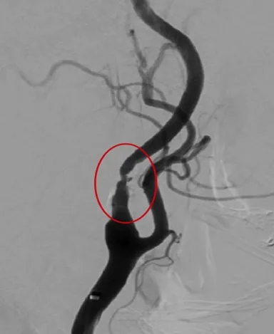 Before stenting
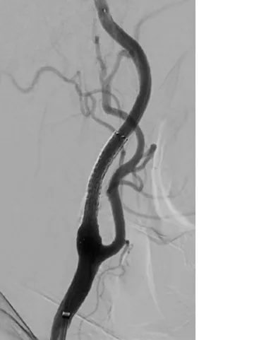 After stenting
A narrowed or blocked vertebral artery can reduce blood flow to the back of the brain. Symptoms may include double vision, slurred speech, poor balance, weakness or numbness, trouble walking, or sudden vision changes. Dizziness alone has many causes. Sudden dizziness with other nerve or brain symptoms needs emergency care.
CT angiography and MR angiography show where the vertebral artery is narrow. They also show whether other vessels can carry enough blood around the blockage. Ultrasound can study parts of these arteries in the neck, but some areas are hard to see. Catheter angiography may be needed if other scans do not explain the symptoms or a procedure is being considered.
Medicine is the foundation of treatment. The goals are to prevent blood clots and slow plaque growth. If symptoms return despite treatment, the team may consider angioplasty, a stent, or surgery in selected cases. The choice depends on the location of disease, alternate blood-flow routes, earlier strokes, and procedure risk.
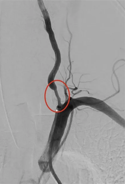 Before treatment
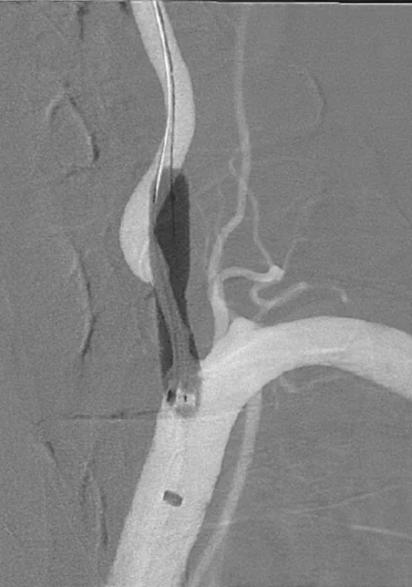 Angioplasty
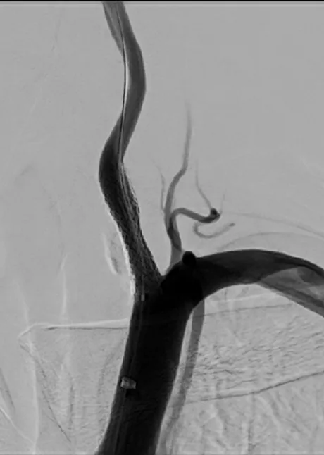 After stenting
Call 911 for sudden facial droop, weakness or numbness on one side, trouble speaking, vision loss, severe imbalance, double vision, or another abrupt change. Symptoms that fade within minutes may still be a TIA. They need urgent care because a full stroke may follow soon afterward.
The degree of narrowing is only one part of the decision. The team asks whether symptoms came from that artery and studies the plaque and nearby vessels. Age, health, earlier treatment, and the likely durability of each option also matter. Together, these factors show who may benefit from a procedure and who is better served by medicine and monitoring.