At the Brain Aneurysm Institute at Beth Israel Deaconess Medical Center in Boston, our specialists review each case together and build a treatment plan around the patient's scans and personal risk. They consider age and overall health, along with the aneurysm's size, shape, location, growth, rupture status, and connection to nearby arteries.
Selected unruptured aneurysms may be monitored with periodic imaging rather than treated immediately. The plan may change if the aneurysm grows, develops a higher-risk shape, causes symptoms, or the patient's health and preferences change.
Microsurgical clipping may be preferred for certain ruptured or unruptured aneurysms. In this procedure, the surgeon places a small titanium clip across the aneurysm neck to stop blood from entering the aneurysm while keeping the main artery open.
Some complex aneurysms involve an artery that cannot be closed safely. Bypass surgery creates another route for blood by connecting a scalp artery or graft to an artery inside the skull.
In this procedure, the surgeon uses a small catheter to guide through the arteries into the aneurysm. Via the catheter, coils are deployed inside the aneurysm sac to promote clotting and reduce blood flow to the region. A possibility of recurrence exists, requiring follow-up imaging to monitor the aneurysm.
Wide-necked or complex aneurysms may need a stent to support the coils and keep the main artery open. In other cases, a temporary balloon helps place the coils and is removed before the procedure ends.
A flow-diverting stent is placed across the aneurysm neck and redirects blood through the main artery. Blood flow inside the aneurysm falls over time, while new tissue grows across the device and repairs the artery from within.
Better scans, surgical methods, and catheter-based devices have expanded the range of aneurysms that can be treated. Even so, not every unruptured aneurysm needs immediate repair. Monitoring, open surgery, and catheter treatment differ in risk, durability, recovery time, and follow-up.
Angiography examples
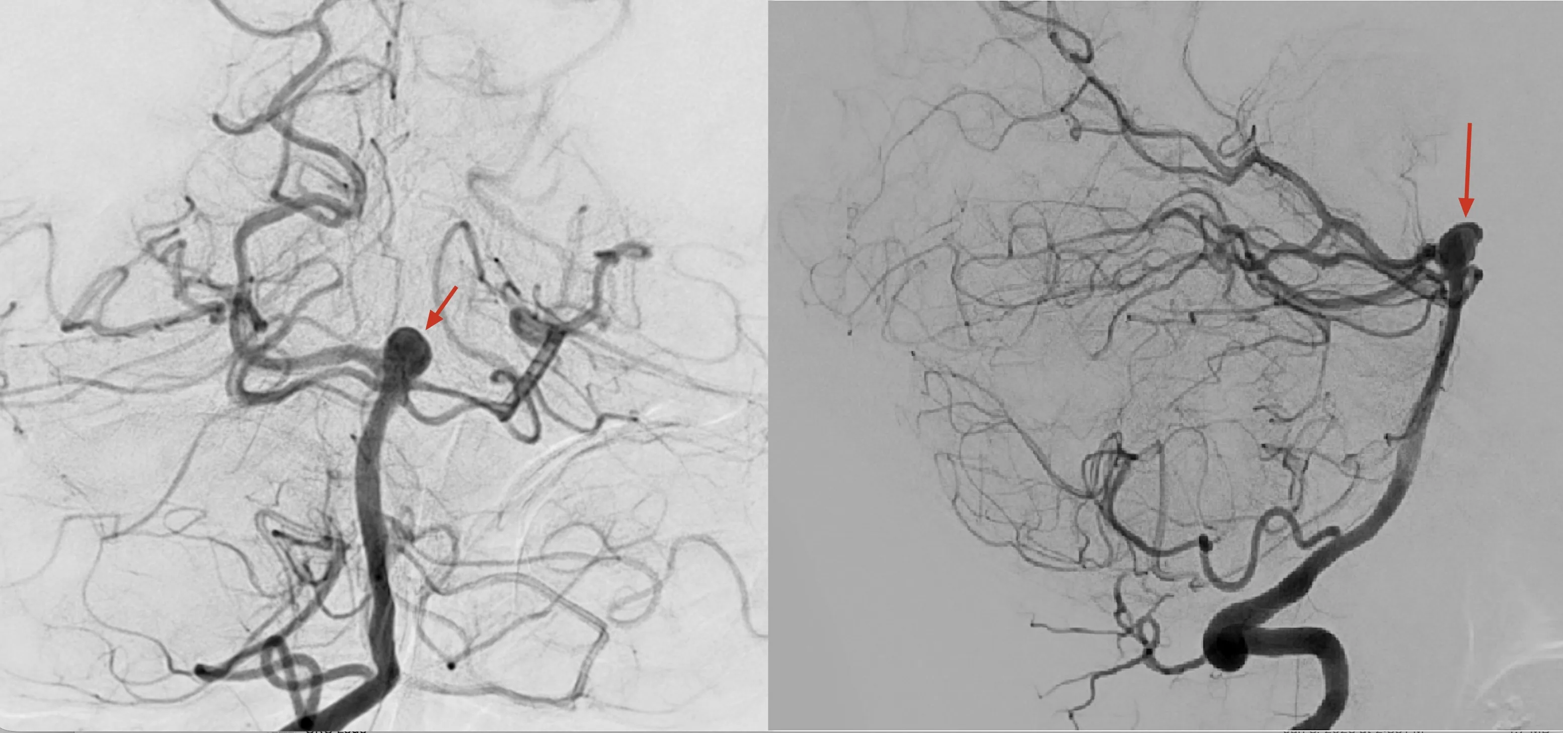
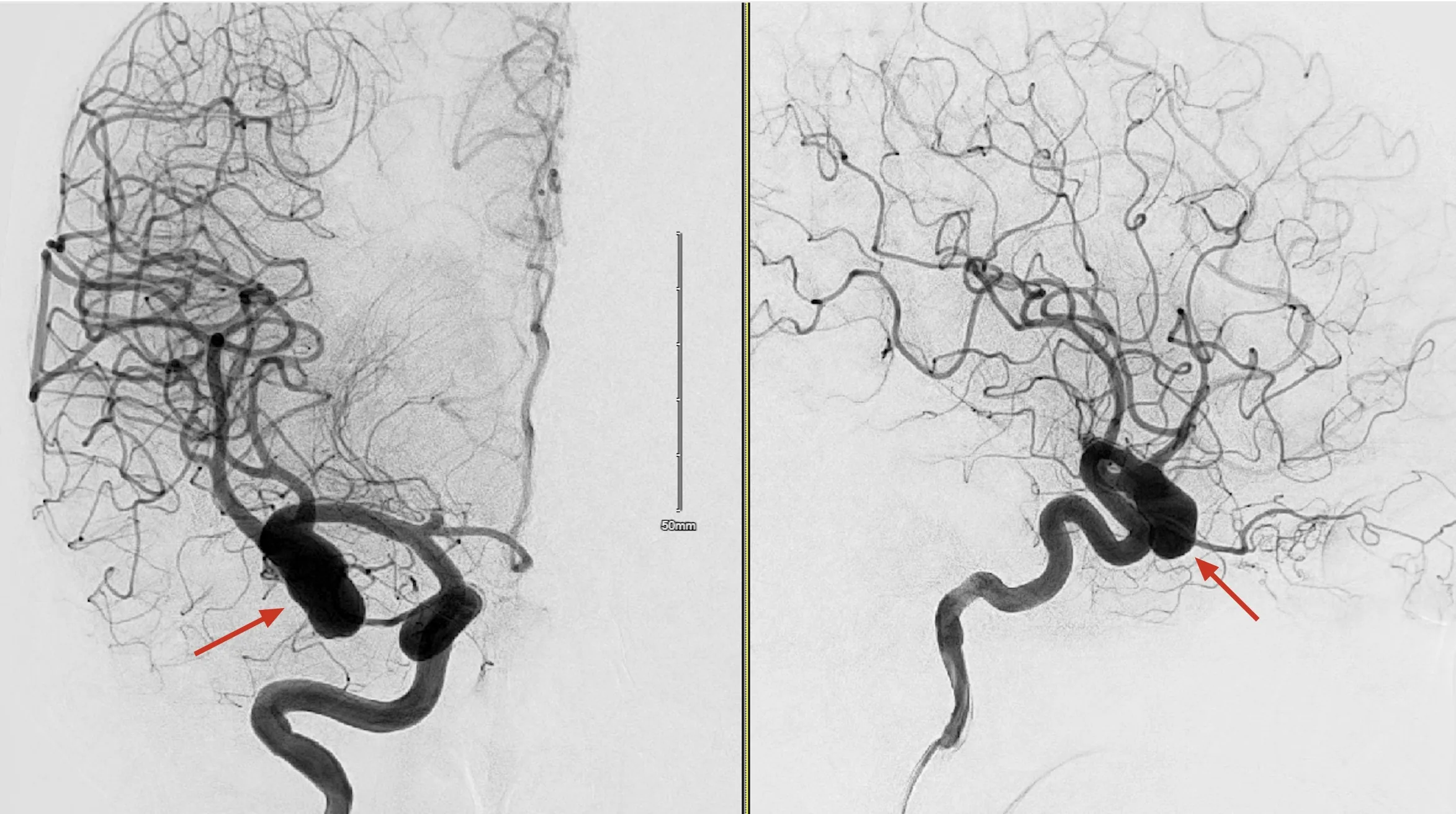
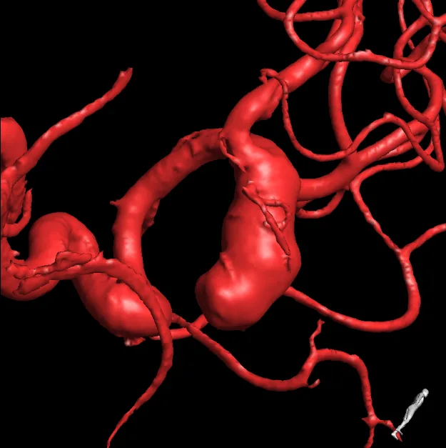
Endovascular treatment reaches the aneurysm from within the arteries, usually through a small opening in the wrist or groin. Doctors guide small catheters with live X-ray images and can treat many vascular disorders without opening the skull. BIDMC's angiography suite has world-class tools and imaging capabilities to visualize the vessels and blood flow in detail throughout the procedure.
The Brain Aneurysm Institute at Beth Israel Deaconess Medical Center, a Harvard Medical School teaching hospital in Boston, offers comprehensive brain aneurysm care. Our team treats aneurysms with both open microsurgical and minimally invasive endovascular techniques. To arrange an evaluation, call 617-632-9940.
Depending on the aneurysm and the patient’s risk, options include monitoring selected unruptured aneurysms with imaging, microsurgical clipping, bypass surgery, endovascular coiling, stent-assisted coiling, and flow diversion. Our specialists match the approach to each aneurysm’s size, shape, location, and rupture status.
The choice depends on the aneurysm’s anatomy, whether it has ruptured, and the patient’s overall health. Because our team is trained in both open and endovascular techniques, we recommend the option best suited to each patient rather than the one dictated by a single specialty.
Yes. Because treatment decisions carry real trade-offs, many patients seek a second opinion at a high-volume center. Our Boston-based team regularly reviews outside imaging and provides second opinions for complex cerebrovascular conditions.