The Brain Aneurysm Institute provides multidisciplinary care for patients with cerebral cavernous malformations, or CCMs. Neurosurgeons, stroke neurologists, seizure neurologists, neuroradiologists, geneticists, rehabilitation specialists, and support staff work together to understand each patient's symptoms, MRI findings, bleeding risk, and treatment options.
Angioma Alliance Center of Excellence Network
Beth Israel Deaconess Medical Center is listed by the Alliance to Cure Cavernous Malformation, formerly Angioma Alliance, as part of its Center of Excellence Network for cavernous malformation care.
Cerebral cavernous malformations (CCMs) are clusters of abnormal, thin-walled blood vessels that can form in the brain or spinal cord. The blood inside them moves slowly and may clot, giving the malformation a rounded, berry-like appearance.
CCMs are not cancer. Many never cause symptoms and are found incidentally on MRI scans. Others can irritate nearby brain tissue, cause seizures, or leak blood, resulting in a symptomatic bleed known as a hemorrhage.
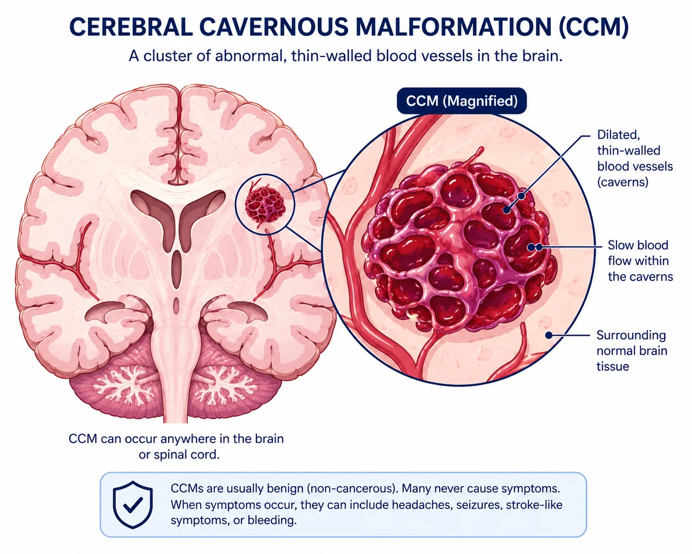
Symptoms depend mostly on where the CCM is located and whether it has bled. A CCM on the outer surface of the brain can irritate the brain and cause seizures. A CCM in the brainstem, deep brain structures, or spinal cord can cause more serious symptoms as those areas are associated with movement, sensation, balance, vision, and other vital functions.
Headaches are common in people with CCMs, but a headache by itself does not always mean a new bleed has occurred. New or worsening neurological symptoms should be discussed urgently with a medical team.
Symptoms can include:
Seek urgent medical care for a first seizure, a seizure that lasts longer than usual, sudden weakness or numbness, trouble speaking, new double vision, severe sudden headache, confusion, loss of consciousness, or other symptoms that could suggest bleeding in the brain or spinal cord.
Magnetic resonance imaging (MRI) is the main diagnostic tool following a CCM. A Computed Tomography (CT) scan can be useful in an emergency when doctors need a fast look for bleeding, but MRI allows much more detail. Specialized MRI sequences can reveal small areas of old blood and help determine presence of multiple CCMs.
Figures 2 and 3 show common MRI appearances. The dark areas come from old blood products. One important finding is a hemosiderin rim: a dark ring made of iron-containing pigment left behind after prior bleeding. In plain terms, it is a footprint of old blood around the malformation.
MRI examples
MRI helps determine where the CCM is, presence of old blood, and how close the malformation is to important brain pathways.
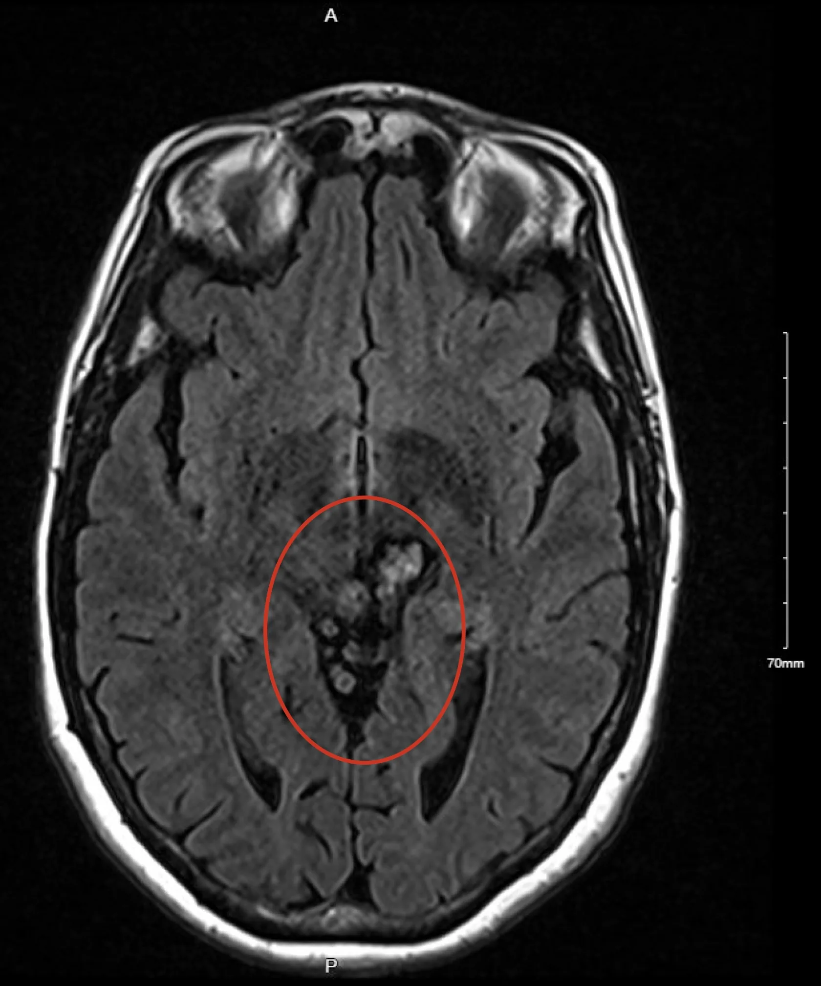
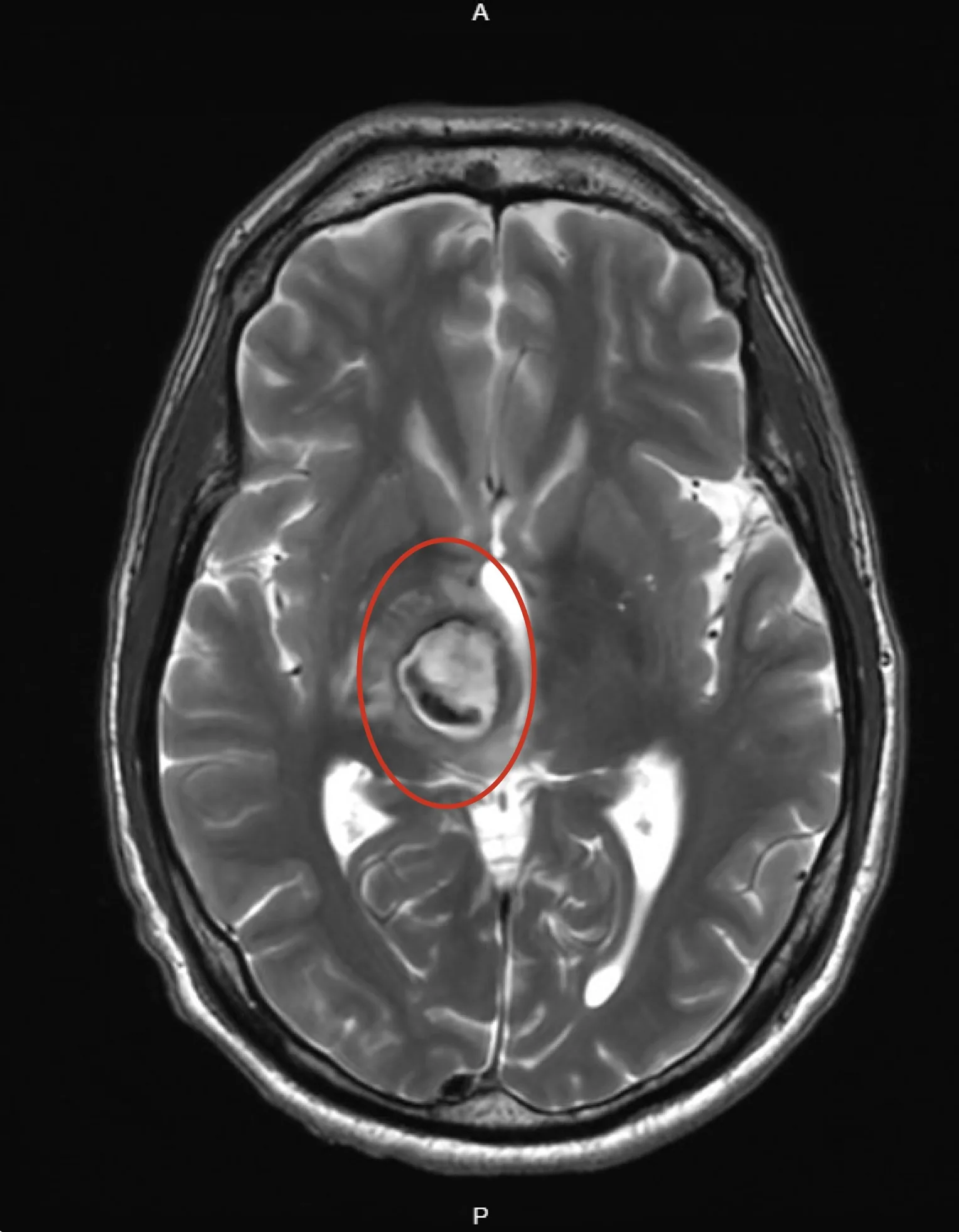
The frequency of follow-up MRI is individualized. Some patients are monitored at regular intervals, while others may only need repeat imaging if new symptoms appear. Cerebral angiography is usually not needed when MRI clearly shows a cavernous malformation, because CCMs are low-flow lesions and often are not visible on standard angiography.
Most CCMs are sporadic, meaning they happen by chance and usually appear as a single malformation. They may occur near a developmental venous anomaly, or DVA, which is an atypical vein pattern that drains normal brain tissue.
Some patients have multiple CCMs. Multiple malformations can be caused by an inherited condition, prior radiation to the brain or spinal cord, or CCM formation near a DVA.
About one-fifth of people with CCMs have an inherited form called familial cavernous malformation syndrome. Genetic testing may be recommended for patients with multiple CCMs without a DVA, or for patients with a family history of CCM.
Familial CCMs are linked to mutations in three genes:
If a hereditary mutation is found, parents, siblings, and children may be offered genetic counseling and testing so they can understand their own risk.
All CCMs can have small amounts of slow leakage over time. A symptomatic hemorrhage is different: it causes new or worsening symptoms because blood irritates or injures nearby brain or spinal cord tissue. The strongest risk factors for another hemorrhage are a prior symptomatic bleed and a CCM located in the brainstem.
Treatment depends on the CCM's location, whether it has bled, whether it is causing seizures or neurological symptoms, and how risky surgery would be for that specific area.
Many incidental CCMs that have never caused symptoms can be watched with clinical follow-up and MRI when appropriate.
Anti-seizure medicines may help control seizures. Other medicines may be reviewed carefully when bleeding risk is part of the decision.
Surgery may be considered after bleeding, for seizures that remain difficult to control, or when the expected benefit outweighs the surgical risk.
Physical, occupational, speech, vision, cognitive, and mental health therapies can help patients recover function and adapt after a bleed or surgery.
A CCM diagnosis can be overwhelming. These questions can help patients and families understand the plan:
Research is ongoing to better understand why CCMs form, why some bleed, and whether future medications can reduce hemorrhage risk or slow the formation of new malformations. At present, surgery is the only treatment that removes a CCM, but many patients never need surgery.
Patients should review activity, pregnancy, blood thinner, hormone therapy, and lifestyle questions with their care team because recommendations depend on the individual situation. In general, maintaining cardiovascular health, avoiding smoking or vaping, avoiding binge drinking, and staying connected to follow-up care are sensible steps.
BIDMC clinicians have participated in natural history studies of cavernous malformations and maintain a close partnership with the Alliance to Cure Cavernous Malformation to support education, research, patients, and families.