The Adult Moyamoya Center at BIDMC brings together experts in stroke care, brain surgery, and imaging. The team studies each patient's arteries, brain blood flow, symptoms, and future stroke risk. These findings guide the choice between medicine, surgery, and continued monitoring.
In neurovasculature, the internal carotid arteries supply the front and middle parts of the brain. Moyamoya disease is a rare disorder in which one or both internal carotid arteries slowly narrow inside the skull. As the disease advances, low blood flow can cause a transient ischemic attack (TIA) or stroke.

The brain responds by growing a network of small vessels known as collateral vessels that carry blood around the narrowed region. On an angiogram, they can resemble a "puff of smoke," giving the disease its Japanese name. The new vessels help, but they may not carry enough blood and can be fragile.
Moyamoya has two common age peaks: one in childhood and another in adulthood. It can affect people of any background, but it is more common among people of East Asian ancestry and may run in families. The team also looks for Moyamoya syndrome, in which the same artery changes occur alongside another medical condition (e.g., down syndrome, neurofibromatosis I, sickle cell disease).
Symptoms can result from low blood flow or bleeding from fragile vessels, with the pattern depending on the part of the brain involved. A TIA causes temporary stroke-like symptoms. An ischemic stroke occurs when low blood flow injures brain tissue. A hemorrhagic stroke occurs when a vessel breaks and bleeds into or around the brain.
Adults may have TIAs, stroke, changes in thinking, seizures, or bleeding in the brain. Even symptoms that go away need prompt evaluation. Moyamoya can progress, and repeated events may cause lasting injury.
Symptoms of reduced brain blood flow or brain hemorrhage may include:
Evaluation may include MRI and MRA, CT angiography, blood-flow studies, and catheter angiography. MRI shows earlier strokes or areas of brain injury. Vascular scans show where the arteries are narrow. Blood-flow testing measures how well the brain responds when demand rises, while angiography maps the collateral vessels for treatment planning.
Direct extracranial-to-intracranial bypass connects a scalp artery to an artery on the brain's surface. The new pathway can improve cerebral blood flow immediately.
EDAS, or encephaloduroarteriosynangiosis, places a scalp artery in contact with the brain's surface. New collateral vessels then grow gradually into the affected region over several months.
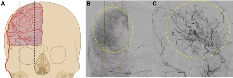
After imaging, stroke specialists and brain surgeons review the findings together. The plan depends on symptoms, disease progress, blood-flow reserve, earlier strokes, and the likely benefit of surgery. Some patients can be followed with medicine and repeat scans. Others benefit from bypass surgery that brings a stronger source of blood to the brain.
Early evaluation can identify patients at risk before another stroke causes lasting injury. Adults with signs of Moyamoya should be assessed by a team experienced in brain blood-flow disorders and bypass surgery.