Vascular malformations and fistulas often need review by several specialists. Our team includes experts in catheter-based treatment, open brain surgery, brain imaging, radiation therapy, and neurology. Care may involve one procedure or a planned series of treatments.
An arteriovenous malformation, or AVM, is an abnormal network where arteries connect directly to veins. The usual bed of tiny vessels between them is missing. This high-flow connection places stress on the vessels and may reduce oxygen delivery to nearby tissue.
AVMs can form in the brain or spinal cord. They may cause bleeding, seizures, headaches, or worsening problems with movement, speech, or sensation. A spinal AVM can also cause pain, weakness, numbness, or changes in walking.
Treatment depends on the AVM's structure, size, location, drainage pattern, symptoms, and history of bleeding. Options may include:
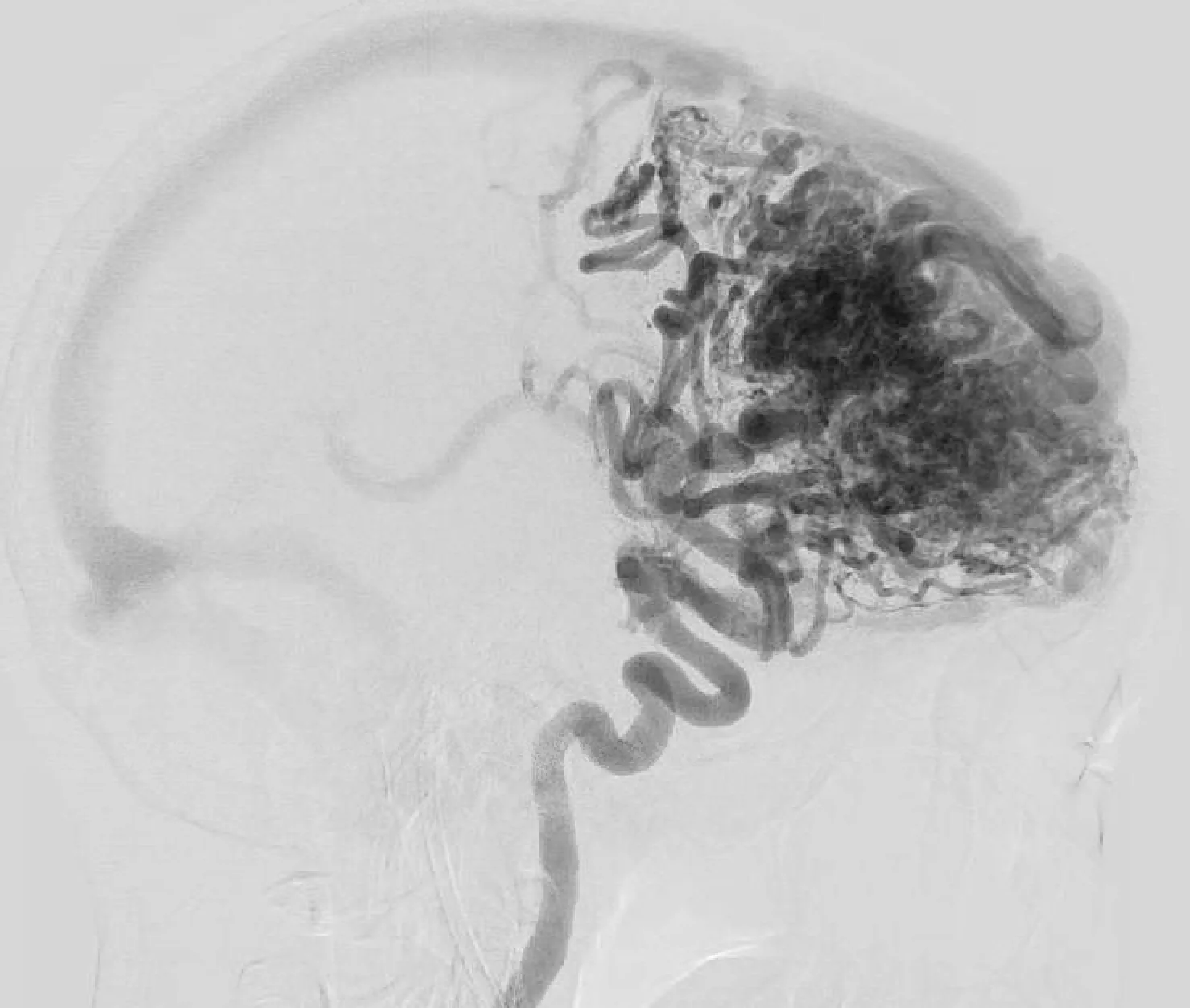 Pre-treatment lateral view
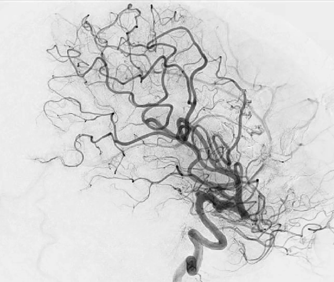 Post-treatment lateral view
A dural arteriovenous fistula, or DAVF, is an abnormal link between arteries in the brain's outer covering and nearby veins. Its risk depends largely on how blood drains from it. Higher-risk fistulas can cause bleeding or lasting injury to the brain or spinal cord.
Treatment closes the abnormal route while protecting normal veins. A catheter-based procedure is often used first. Surgery or focused radiation may suit other forms of the condition.
A carotid-cavernous fistula, or CCF, is an abnormal connection between a carotid artery and a vein-filled space behind the eye. Rising pressure in this area can cause eye redness, swelling, pulsing, double vision, or loss of vision.
Catheter-based treatment can close the fistula and reduce pressure behind the eye. The goal is to protect vision and keep normal blood flow through the carotid artery.
Carotid body tumors are growths with a rich blood supply. They form near the point where the carotid artery divides in the neck. Brain, vascular, and head and neck surgeons may work together because the tumor lies close to major arteries and nerves.
Embolization may reduce blood flow to the tumor before surgery. In some cases, the team may recommend radiation or careful observation based on growth, symptoms, and treatment risk.
Before recommending care, the team maps the arteries feeding the lesion, the veins draining it, and nearby parts of the brain. This review shows which vessels can be treated safely and whether treatment should occur in stages.